5 Questions You May Have About Your Retinal Consultation
What is a retina specialist?
A retina specialist is a highly trained medical professional who specializes in the treatment of conditions and diseases affecting the retina. Your doctor may refer to you a retina specialist for a consultation if they notice early signs of retinal disease or if they are concerned you may develop it in the future. Under the care of a retina specialist, it is possible for patients suffering from retinal conditions to restore lost vision and slow or prevent future damage.
What does a retinal consultation look like?
A retinal consultation begins in a very similar manner as a routine vision exam. An ophthalmic technician will take your medical history, evaluate your vision in order to determine how well you are seeing, and test your eye pressure levels. Once this is completed, your eyes will be dilated. Dilating your eyes allows for a clear view of the back of your eye, including your retina. It takes 20 to 30 minutes for your pupil to fully dilate.
What kinds of tests or imaging will be used to examine my eyes?
While your eyes are dilating, the doctor may order specialized testing to be performed. These tests may include:
Optical Coherence Tomography or OCT
An OCT test is a non-invasive imaging test that uses light waves to take cross-section pictures of your retina. With these images, the retina specialist can see the layers of your retina and measure their thickness. This information helps the retina specialist determine if you are suffering from any retinal diseases. The test itself requires you to sit in front of the OCT machine and remain motionless. At no time will the equipment touch your eye, however you may be light sensitive for several hours after the test.
Fluorescein Angiography
Fluorescein angiography is a diagnostic test that utilizes a camera to record the blood flow through your retina. This is performed by injecting a fluorescein dye into your arm or hand. Once the dye is injected, a series of photographs will be taken of your eye in order to document how blood is traveling through your retina. The pictures of your retina are in black and white and the dye appears white or gray, illuminating the blood vessels for a clearer photo. The photos produced by this test allow the retina specialist to see if your blood vessels are leaking, how much blood is leaking, and where the leaking is occurring. At no time will the equipment touch your eye, however some individuals may experience nausea during the procedure, which usually subsides quickly.
Fundus Photography
Using a fundus camera, a highly specialized camera, pictures are taken of the structures of the back of the eye. These pictures are used to document and diagnose certain eye conditions. At no time will the equipment touch your eye, however you may be light sensitive for several hours after the test.
Fundus Autofluorescence Imaging
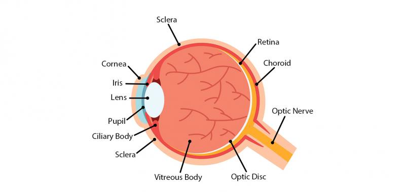
Through the use of autofluorescence imaging, the doctor can observe your eye’s fundus, the eye’s interior surface that sits opposite the lens and includes the retina, macula, fovea, optic disc, and posterior pole. By observing the fundus, retinal specialists can detect eye diseases. This is a non-invasive test and at no time will the equipment touch your eye.
How will the doctor determine my treatment options?
Once testing is complete and your eyes are fully dilated, the doctor will see you. You will be asked to describe any symptoms you are experiencing.
The doctor will then examine the structures of the back of your eye. Because of this, there may be times in which the doctor has to shine a bright light into your eye and manipulate your eye to get a clear view. Although this may be uncomfortable, it is an important step in the process and your doctor will complete it as quickly as possible.
Your doctor may use a slit lamp, a specialized microscope, to aid in this process. They also an indirect ophthalmoscope to get a broad view of the retina.
After a thorough review of your retina and the results of any testing you received, the doctor will discuss your diagnosis and possible treatment path. Depending on the diagnosis, your treatment may begin immediately or you may be watched for further development. Although you will receive a thorough exam, you will not leave the office with a prescription for glasses or contacts.
How can I prepare for my appointment?
To prepare for your retina consultation, you should compile a list of the names of any medications you may be taking as well as a complete medical history. Due to the complex nature of the consult, you should plan on being at the office for a minimum of two hours.